


The Human Knee: Anatomy, Motion, and How It Works
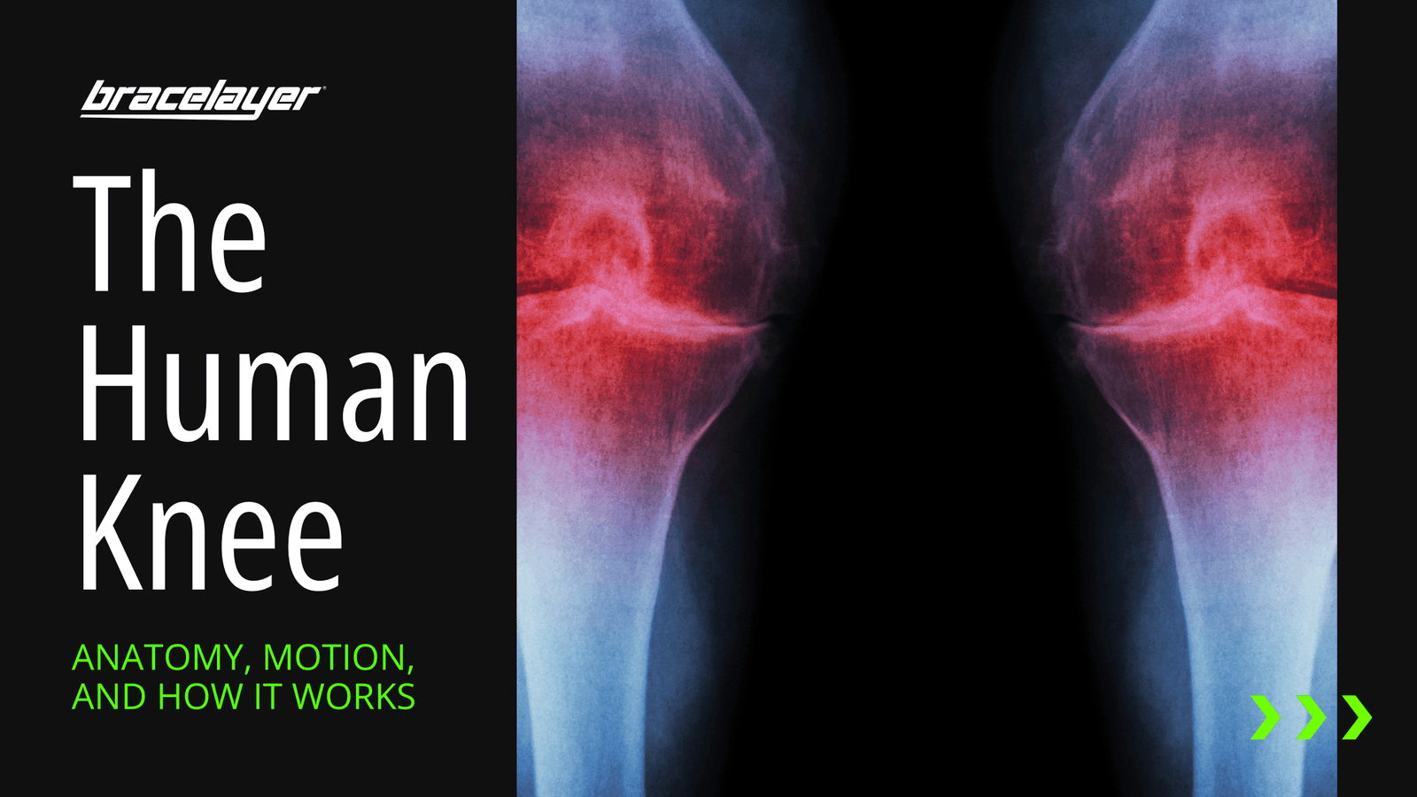
The knee is one of the largest and most important joints in the human body, tasked with the important job of connecting the upper leg (femur) with the lower leg (tibia or shinbone) to facilitate motion. At its most simplistic definition, one may consider the knee to simply be two leg bones joined together by muscles, ligaments, and tendons. But the knee is much more than the sum of its parts. Indeed, it’s the precise, nimble, effortless way that the joint’s composition works in unison to create movement that makes the knee such a crucial component of the human anatomy.
So, let’s start at the beginning. When talking about the knee, one should start by learning the basic components of the joint.
The knee is composed of three main structures:
- The tibia is the shin bone, the largest bone of the lower part of the leg
- The femur is the thigh bone, part of the upper leg bone
- The patella is also referred to as the kneecap
What causes the knee to move?
The muscles that propel the knee’s natural movement and rotation are connected by tendons to the knee bones. Tendons are bands of connective tissue on the end of muscles, connecting muscle to bone. Muscles and tendons work in unison. The muscle contracts and the tendon’s tough, cordlike tissue, which is connecting that muscle to the bone is pulled, causes the bone to move.
- The quadriceps muscles work in conjunction with quadriceps tendons to straighten (extend) the knee. They provide strength and power to create the knee’s extension.
- The hamstrings muscles allow for flexion (bending)
Ligaments and tendons
In addition to muscle and bones, the knee joint is comprised of tendons and ligaments, two similar yet slightly different types of connective tissue in the body. How do they differ?
Ligaments are bands of connective tissue with collagen fibers, which connect knee bones to each other and help to provide stability. Tendons specifically connect muscle to bone.
Tendons and ligaments also vary in other ways. Ligaments crisscross in direction, as they attach bone to bone to stabilize joints. Since they help with movement, tendons have a bit more elasticity than ligaments. Ligaments are tight and help to stabilize the knee.
There are several types of tendons in the knee including the patellar tendon, which attaches the tibia to the patella. The quadriceps tendon attaches the quadriceps muscle to the patella. Many types of ligaments and tendons can be found throughout the body.
Other components of the knee
The knee also contains menisci and cartilage which function as shock absorbers, allowing the knee to move with less friction.
- Articular cartilage is the smooth lining covering the end of the bone. If it’s damaged or worn away, an individual may develop arthritis of the knee.
- Meniscus: is contained in the area between the end of the thigh bone and the top of the shin bone. The meniscus help distribute the body’s weight evenly, adding balance and stability.
There is also the joint bursa (synovial bursa), a closed fluid filled sac in front of the knee, just below the surface of the skin. It cushions the bones, tendons, and muscles. It smooths out motion between the skin and bone, with the fluid allowing muscles and tendons to glide over ligaments and/or bone.
There are various bursae in the knee performing this function, including the pre-patellar bursa. And there are hundreds of bursae throughout the body, each acting in a similar capacity for various parts of the anatomy.
Two fun facts about the knees
Fact 1: Surprisingly, our kneecaps don’t harden until somewhere between the ages of 3 to 6. As noted by Healthline this may be because toddlers are still learning to walk, and the softness of a still undeveloped knee may provide extra cushioning for all those falls. It may also be to make the birth process easier. They do have cartilage that will eventually become part of the kneecap. But it’s not fully formed until later in childhood when it begins to ossify or turn into bone.
Fact 2: According to the UK Readers Digest, the patella (kneecap) is a type of sesamoid bone, a bone embedded within the tendon. In the case of the patella, the bone is within the quadriceps tendon. They differ from other bones, which are connected to each other externally, instead of being embedded in tendon. Other sesamoid bones are found in the feet and hands.

A little bit about knee pain
Knee pain is quite common, often associated with aging or daily wear and tear from walking, standing, bending, lifting, etc. People who participate in more athletic activities that involve motions such as pivoting, jumping, or running, whether professional or amateur, are also likely to suffer from injuries or conditions involving the knee.
The most common knee conditions
Torn meniscus (meniscal tear): One common injury is a torn meniscus. This condition is often caused by forcefully twisting or rotating your knee, especially when your full weight is on the joint.
Hyperextension: This occurs when the joint’s angle is stretched, straightened or opened beyond its normal, healthy range of motion. This can cause tears to the tendons or ligaments or cartilage damage. A sprained knee is one common type of hyperextension injury.
ACL tear: Because the anterior cruciate ligament (ACL) helps give the knee stability, tears of the ACL often makes the individual feel as if their knee is “giving out.” It is a ligament that stretches through the middle of the knee to keep the tibia in place.
Knee bursitis: This is inflammation, swelling, or pain usually caused by overuse or injury.
Exercise helps knee health
It is encouraged that people walk briskly or do other light to moderate physical exercise on a regular basis to maintain the condition of joints and limbs, as well as to ensure their overall health.
It is commonly suggested that you do stretching exercises before and after workouts or other physical activities to prevent injuries such as these. You should always contact a doctor before beginning any new workouts that may be strenuous.
Check out our infographic below for a visual breakdown of the anatomy of your knee:

Leave a comment